





The retina lifts
The retina is the thin, clear layer at the back of the eye that works like camera film. In a detachment, its inner layers and light-sensing cells pull away from the retinal pigment epithelium beneath them.
An eye emergency only you can see
Retinal detachment happens when the retina peels away from the inner wall of the eye. If it spreads to the macula, blindness can follow, so it is treated as an emergency. At Sky Eye Clinic in Seoul, the retina team examines you and explains laser, vitrectomy, scleral buckling and gas options.
No one can see a detachment by looking at you. Only you feel the signs, so act on them quickly.
The retina is the thin, clear layer at the back of the eye that works like camera film. In a detachment, its inner layers and light-sensing cells pull away from the retinal pigment epithelium beneath them.
Once the detachment reaches the macula at the center, nourishment and blood supply fail and sight can be lost. That is why speed matters.
Detachment is no longer only a problem of older eyes; younger people now get it often as well. High myopia calls for particular care.
A tear or hole forms through injury, surgery, high myopia or age. Vitreous fluid slips through it and lifts the retina.
Membranes left by diabetic retinopathy, uveitis, high blood pressure, retinal vasculitis or retinopathy of prematurity pull the retina away.
Fluid gathers under the retina because of retinal or choroidal disease. It spreads slowly from the edges, rarely causes symptoms and tends to be discovered incidentally.
A detachment caught at an early, limited stage is treated straight away with laser photocoagulation. Once it has progressed, one of three operations is needed.
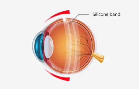
A silicone band is fixed to the sclera, the outer shell of the eye, and tightened so it presses the retina back into place. When the tear closes, the fluid beneath is absorbed and the retina settles where it belongs.
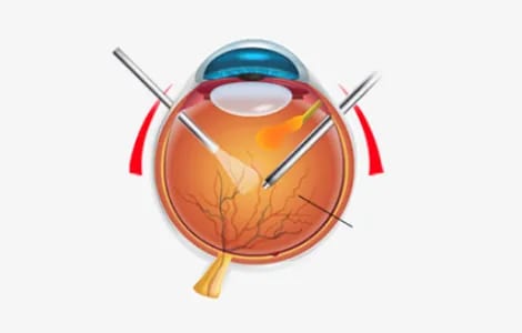
The surgeon works through tiny openings, takes out the vitreous gel and fills the space with gas or oil that presses the retina flat.
The retina returns to position faster, but you need to hold a set head position for a period while it heals.
A large bubble of gas is placed in the middle of the eye. Its main uses are tears in the upper outer retina, macular holes, a retina that detaches again after scleral buckling, and bleeding.
As with vitrectomy, you rest in a set position for a period afterwards.
Which method suits you depends on the type of detachment and how far it has spread, and recovery differs from eye to eye. If gas or oil is used, the head position you keep afterwards is part of the treatment.
Detachment can follow diabetic retinopathy or uveitis. A different membrane problem is explained on the epiretinal membrane page.
New floaters, flashes, a shadow like a curtain over your view, or sight that suddenly drops. Any one of them is a reason to be examined quickly.
High myopia, age, earlier eye surgery, retinal problems in the family and injury to the eye all raise the risk.
When it is caught while still small and limited, it is sealed with laser photocoagulation straight away.
Yes, after vitrectomy or pneumatic retinopexy you rest in a specific position for a period so the retina can heal.