





The optic nerve
The optic nerve carries what you see to the brain. When high pressure or poor blood flow injures it, your visual field closes in gradually.
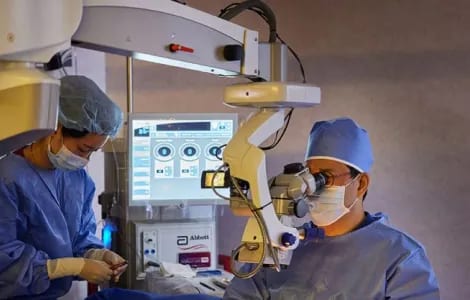
Your field of view can narrow without a single symptom
Glaucoma is damage to the optic nerve, usually from high eye pressure or poor blood flow, that slowly shrinks the area you can see. At Sky Eye Clinic in Seoul, doctors measure your eye pressure, identify the type and explain drops, laser and surgery.
Early glaucoma rarely gives clear symptoms. Regular eye exams are how it is found in time.
The optic nerve carries what you see to the brain. When high pressure or poor blood flow injures it, your visual field closes in gradually.
A small amount of fluid, the aqueous humor, nourishes the cornea and lens. If its outlet narrows or blocks, pressure builds and the pressure-sensitive nerve is damaged. Severe cases can end in blindness.
The exact cause is not fully understood. High eye pressure is the main factor, and reduced blood supply to the eye also plays a part in how it begins and progresses.
Early glaucoma has no clear symptoms; they arrive late. By then the field of view has narrowed a great deal, which makes daily life harder and slows your reaction in an emergency.
The most common type. The drainage outlet wears down gradually and the disease moves slowly.
Pressure is high, yet the eye looks normal and nothing feels wrong while sight narrows.
Usually sudden. The drainage path blocks all at once and pressure climbs fast, bringing pain, a red eye, headache, nausea and rings around lights.
A form of open-angle glaucoma in which pressure reads normal, but the optic nerve shows typical changes and the visual field is lost. Only regular exams catch it early. A family history or heart problems such as arrhythmia make it more likely.
Triggered by another eye or general health problem, for example injury, inflammation, a tumor, bleeding, previous surgery, a cataract left for a long time, developmental abnormalities or steroid use.
The drainage path failed to develop normally before birth. Very large-looking eyes or constant tearing in a newborn are reasons to suspect it.
Medication is the basic treatment, and its main goal is to lower eye pressure. Your doctor chooses drops by your condition and response, then checks at follow-ups that they are working.
There are now more kinds of drops, and they lower pressure better, so for many patients drops are all that is needed.
In angle-closure glaucoma, laser iridotomy opens a small hole in the iris and can control pressure much faster than medicine.
When medication is difficult, trabeculoplasty treats the trabecular meshwork, the eye’s drainage tissue, so fluid can flow out more easily.
If drops or laser do not bring pressure down enough, surgery creates a route for fluid to drain under the conjunctiva, the thin layer over the white of the eye.
Options include trabeculectomy and the implantation of an aqueous shunt.

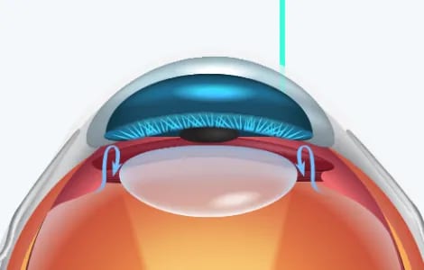
Glaucoma care sits within our Retina Center, so your plan is made alongside what your retinal exam shows. How well pressure responds to each step varies from person to person, which is why follow-up visits are part of treatment.
Glaucoma can also develop after uveitis or retinal vein occlusion.
Usually not. Symptoms tend to appear only late, once the field of view has already narrowed. Regular exams are how it is caught early.
Raised eye pressure, glaucoma in the family, being 40 or older, myopia, diabetes (4 times the risk) and high blood pressure (2 times the risk).
It tends to come on suddenly: a painful red eye, headache, nausea and halos around lights. Treat it as urgent.
For many people, yes. The aim is lower eye pressure, and modern drops achieve it more effectively. If they fall short, laser or surgery follows.