





Arteries and veins of the retina
Arteries bring blood into the retina, veins carry used blood back toward the heart, and both branch out. Vision lost because one of these vessels has closed off is called retinal vascular occlusion.
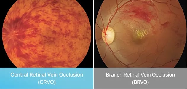
When a retinal vessel blocks, hours count
Retinal vascular disease covers problems with the blood vessels of the retina, most often an artery or vein that becomes blocked. A blocked artery can take sight suddenly; a blocked vein usually blurs it. At Sky Eye Clinic in Seoul, the retina team examines your eye and explains the treatment for each.
For a blocked artery, the aim is to get blood flowing again within the first 24 hours.
Arteries bring blood into the retina, veins carry used blood back toward the heart, and both branch out. Vision lost because one of these vessels has closed off is called retinal vascular occlusion.
An occlusion is named by where it is and how much it covers: central or branch retinal artery occlusion, and central or branch retinal vein occlusion.
The group also includes ocular ischemic syndrome and hypertensive retinopathy. Many of these problems are tied to diabetes and blood pressure.
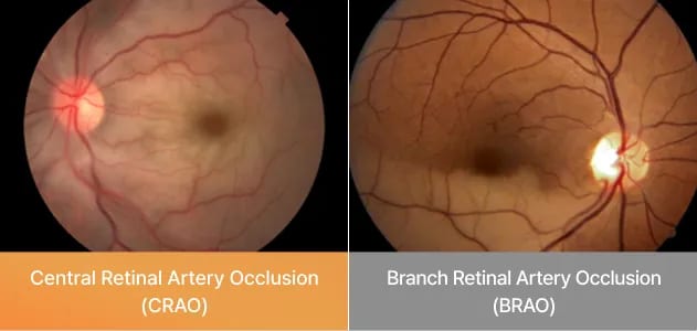
The eye depends on blood from the choroid and the central retinal artery for oxygen and nutrients. A blockage cuts off that supply. It mostly affects people whose circulation is impaired by diabetes, obesity, high blood pressure or heart disease, and adults aged 50 to 70.
It tends to occur alongside arteriosclerosis, high blood pressure, diabetes and high blood lipids, all of which disturb circulation. Other causes are possible too.
Vision drops suddenly or goes dark for a while. It usually affects one eye; both are involved in about 1 to 2% of cases.
Sight becomes dimmer and blurred, also usually in one eye. It can trigger new vessel growth and complications including glaucoma, which is why it needs regular follow-up.
Blood flow to a blocked artery needs to be restored within 24 hours. The longer it waits, the harder it is to win vision back.
The aim is to get blood moving again within 24 hours. That includes bringing eye pressure down quickly and massaging the eye.
Medicines that widen the vessels (vasodilators) or prevent clotting (anticoagulants) may be added, depending on your condition.
Retinal swelling is brought down with laser, an anti-VEGF injection or a steroid injection.
Some eyes also receive fundus photocoagulation to keep new vessels from forming.
How much vision returns depends heavily on how soon treatment starts, and it differs from person to person. Related conditions are explained on the diabetic retinopathy and glaucoma pages.
A sudden loss of vision or a spell of temporary blindness, usually in one eye. Both eyes are affected in about 1 to 2% of cases.
Blood flow should be restored within 24 hours. Waiting longer can make vision recovery difficult.
Swelling is treated with laser, anti-VEGF or steroid injections, and fundus photocoagulation may be used against new vessel growth.
People with diabetes, obesity, high blood pressure, heart disease, arteriosclerosis or high blood lipids, and adults aged 50 to 70.